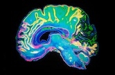
HYPNOSIS AND NEUROSCIENCES
Interacting brains.

Subdivision of human neocortex.
Taken and Modified from http://serendip.brynmawr.edu/bb/
Such expansion allowed the emergence of fundamental abilities such as verbal language, an unique characteristic of human beings, that has its roots in non verbal skills already present in primates.
Our species evolved and populated a world inhabited mainly by other human beings, that is, a world in which social interaction is fundamental. Verbal (and non verbal) communication therefore represented the basis of effective hunting, of the creation of alliances and of the reciprocal exchange of knowledge, processes that were crucial for the development of societies.
Before speaking of hypnotic induction, it is important to consider that in everyday life we are able to modify our mental states, on the basis of what is said by other people, and therefore to modify mental states of others. From this point of view, natural hypnosis as described by M. Erickson presents itself as a direct functional derivation of language itself.
The brain in the mirror
The discovery of mirror neurons further supported this hypothesis. Through studies conducted at first on monkeys and then on men, it was demonstrated that in frontal brain areas specific motor neurons, which activate during the execution of a movement, also react to the view of another human performing that same movement. But there’s more. Some of these neurons simply activate in the observer when the intention of movement of the observed subject is recognized: for instance when, looking at a table that has to be cleared, I see an hand close to the dishes and I hypothesize, unconsciously, the action that is going to be executed. Further research have identified different brain areas that are part of the “mirror circuit” (figure below).

Human mirror circuit.
Prefrontal areas are in green, parietal areas are yellow and the temporal superior sulcus is red.
This new definition of mirror neurons, while representing an interesting interpretation of the development of language, made researchers wonder about the idea of empathy. Understanding others’ emotions may, in fact, be explained through a neural mechanism of such kind that may make the decoding immediate, needless of any conscious effort.
Given this innate cerebral skill for decoding others’ mental states and for influencing them through language and expressions, it is natural to ask ourselves how wide our area of influence is. Maybe the answer is: it depends on the context. It would be unthinkable to say to a child who just fell “stop feeling pain”, expecting to instantly sedate all his tears. During the ‘40s scientists already observed that soldiers who fought during World War II, were able to walk for long distances while they were in danger because of enemy’s fire, even when they were heavily wounded. War environment made their brain forget about physical pain by activating endogen opioid circuits (which regulate the modulation of pain), to preserve a far more precious thing: their lives. Even without referring to such extreme situations, some clear examples of how the context influences ongoing therapies can be found in literature. The fundamental context that can be found in therapy is represented by the relationship between the therapist himself (may he be a physician, a physiotherapist or a psychologist) and his patients.
Hypnotic induction and the brain
Hypnosis, as an instrument used in therapeutic relationships, has been explored since the early ‘60s through electroencephalography (EEG). EEG is a non-invasive technique that detects extra-cortical spontaneous activity. Observing the main brain rhythms (figure below), represented by alpha, theta and delta waves, hypnosis could emancipate itself from the simple definition of being a sleep-like condition.

The main brain wave types of the sleep-wake cycle.
Some possible markers of the hypnotic state have been found in specific wave groups that are absent during deep sleep. For instance, since long time, the correlation between higher presence of alpha waves in both hemispheres and the tendency to suggestibility has been confirmed, suggesting that this rhythm can be a cue that indicates the cognitive style of people who are skillful in having hypnotic experiences. An interesting finding demonstrates that the right parietal-temporal region of the most suggestible subjects shows more electric activity compared to the left side, as opposed to less suggestible subjects. Still, using indirect Ericksonian hypnotic inductions, the same higher right activation can be detected in low suggestible subjects, highlighting how important the associative parietal-temporal area is in hypnotic induction.
The most recent studies on neuroanatomical correlates of hypnosis use modern brain imaging techniques based on blood flow (fMRI – Functional Magnetic Resonance -, and PET – Positron Emission Tomography - ). These techniques work on the assumption that most active areas will receive more blood than the others. In a recent experiment, it has been showed that it is possible to modulate pain through hypnosis in fibromyalgia patients. Results showed that, after a hypnotic analgesic suggestion, a subjective perception of pain reduction was accompanied by high activation of many areas such as the anterior cingulate cortex, posterior and anterior insula, cerebellum and parietal inferior cortex (see image below). Further studies examined the importance, during hypnotic induction, of the corpus callosum, that is the brain structure primarily responsible of the exchange of information between hemispheres. The rostral anterior part of the corpus callosum, in fact, connects the prefrontal cortex of both hemispheres. These areas are generally involved in shifting attention, supporting the hypothesis that hypnotic states are explainable as transient modifications of the subject’s attention (a shift from the inside to the outside).

Activated areas during hypnosis induced analgesia.
From left to right: areas that are activated in hypnotized subjects are in the first column; areas of subjects without hypnotic induction are in the second column, and in the third one, the differences between such areas. From Derbyshire et al. (2008).
Alessandro Piedimonte
For a more thorough explanation of neurosciences and hypnosis see the appendix of the book “Hypnosis and Palliative Care”.
Bibliography
1. Kaas J.H.: The evolution of the complex sensory and motor systems of the human
brain. Brain Res. Bullettin, 75, 384 – 390, (2008).
2. Chomsky N.: Language and the mind. Psychol. Today, 1 (9), 48 -68, (1968).
3. Rizzolatti G., Craighero L.: The Mirror-Neuron System. Annual
Rev. Neurosci., 27, 169 - 92, (2004).
4. Iacoboni, M., Molnar-Szakacs, I., Gallese, V., Buccino, G., Mazziotta, J.C.,
Rizzolatti, G.: Grasping the intentions of others with one's own mirror neuron
system. PLoS Biology, 3 (3), e79, (2005).
5. Del Castello E., De Benedittis G., Valerio C.: Dall'Ipnosi Ericksoniana alle
Neouroscienze. Franco Angeli Editore, (2008).
6. Beecher H. K.: Relationship of significance of wound to pain experienced.
J Am Med Assoc. 161 (17), 1609-13, (1956).
7. Dobrilla G.: Placebo e dintorni. Il Pensiero Scientifico Editore, (2004).
8. Morgan H. A., Macdonald H., Hilgard E. R.: EEG Alpha: Lateral Asymmetry
Related to Task, and Hypnotizability. Psychophysiology, 11 (3), 275 – 282, (1974).
9. Mészáros I, Szabó C. Correlation of EEG asymmetry and hypnotic susceptibility.
Acta Physiol. Hung., 86(3-4), 259-63 (1999).
10. Derbyshire S. W. G., Whalley M. G., Oakley D. A.: Fibromyalgia pain and its
modulation by hypnotic and non-hypnotic suggestion: An fMRI analysis.
European Journal of Pain, 13, 542 - 50, (2009).
11. Horton J. E., Crawford H. J., Harrington G., Downs J. D.: Increased anterior
corpus callousum size associated positively with hypnotizability and the ability
to control pain.
Brain: A Journal of Neurology, 127, 1741 – 1747 (2004).
12. Gruzelier J. H. Frontal functions, connectivity and neural efficiency underpinning
hypnosis and hypnotic susceptibility. Contemporary Hypnosis, 23 (1), 15-32, (2006).
Links
[ NEUROPHYCHOLOGY ] www.neuropsicologia.it
An Italian neuropsychology website in which interesting information about the history, methods and the most common neuropsychological tests can be found.
Site in Italian.
[NEUROPSY] www.neuropsy.it.
A well organized neuropsychology website about pathologies that can alter and reduce cognitive skills of human brain. Its blog is very interesting (http://www.neuropsy.it/blog/) and contains many interesting articles about neurosciences and general psychology.
Site in Italian.
[ NEUROSCIENCES ] www.neuroscienze.net.
An online magazine entirely dedicated to everything that concerns man and his brain. In the section “tematiche” different areas can be reached such as, for instance, neurosciences, psychology or cognitive sciences. It is regularly updated with new articles.
Site in Italian.
[ NEUROSCIENCE FOR KIDS ] www.faculty.washington.edu/chudler/introb.html
Despite its name, it’s a well built website that allows to learn the basic notions about the nervous system. Furthermore, it has an amusing section called “experiment” in which nice games connected to cognitive functions can be played. Site in English.
[ THE BRAIN FROM TOP TO BOTTOM ] www.thebrain.mcgill.ca
Certainly one of the best popular scientific websites concerning the sciences that study the brain and cognition. Clicking on any topic you can access a page where you can choose the level of explanations (socially, psychologically and molecularly).
Site in English.

